


Spectral Optical Coherence Tomography (S-OCT)
[sliders] [slider title=”What is it?”] What exactly is the spectral optical coherence tomography? The spectral optical coherence tomography (Spectral Optical Coherence Tomography: OCT-S) is the newest imaging method of the macula and the optic nerve. It is a cutting edge technology examination through which avery detailed mapping of the posterior eye structures takes place. https://www.youtube.com/watch?v=FjtZIhHbheE&w=600&h=420 […]
[sliders]
[slider title=”What is it?”]
What exactly is the spectral optical coherence tomography?
The spectral optical coherence tomography (Spectral Optical Coherence Tomography: OCT-S) is the newest imaging method of the macula and the optic nerve. It is a cutting edge technology examination through which avery detailed mapping of the posterior eye structures takes place.
https://www.youtube.com/watch?v=FjtZIhHbheE&w=600&h=420
Optical coherence tomography (OCT) with the system Heidelberg Spectralis:
Through the OCT examination, it is accomplished, in real time, a full tomographic estimation (optical biopsy), of both the structure and pathology of the suffering area (cases of diabetic retinopathy, age-related macular degeneration, other maculopathy, hole kystoeidous edema macula, central serous chorioeidoamfivlistroeidopatheias, glaucoma, edema central region, etc. may be diagnosed). The OCT diagnostic technique is performed by scanning the point we want to check with a special laser.
 13")
 14")
The findings are analyzed by the software of the machine and an image that is displayed on the screen is created. This image is ultimately a visual intersection of the point of interest, which allows us to estimate, with certainty, the type of failure and adapt our therapeutic practice accordingly. Note that the method, in addition to its diagnostic value, is of particular importance to monitoring the evolution of eye diseases (follow-up) for any changes in treatment.
https://www.youtube.com/watch?v=WDkHWVGwxQ&w=600&h=420
[/slider][slider title=”Procedure”]
What is the procedure in the OCT examination (optical coherence tomography)?
The test requires no special preparation of the patient’s side. The patient is placed in front of the machine (with a canopy mirror) and within the minimum amount of time, a detailed recording (mapping) of macula takes place . The time required for the recording is just a dew seconds.
 15")
 16")
https://www.youtube.com/watch?v=OgUfCQG3ztE&w=600&h=420
It should be pointed out that an optical coherence tomography machine does not use “dangerous” radiation. Its application, even on a regular basis, is absolutely secure.
https://www.youtube.com/watch?v=w4vFoSIyWs&w=600&h=420
[/slider][/sliders]
Απόφραξη Φλέβας Αμφιβληστροειδή: «Οφθαλμικό εγκεφαλικό»
Τι ακριβώς είναι η απόφραξη φλέβας αμφιβληστροειδή; Φλεβική απόφραξη συμβαίνει όταν στην πραγματικότητα αποκλειστεί μια φλέβα στο οπίσθιο τμήμα του οφθαλμού, δηλαδή στον φωτοευαίσθητο χιτώνα που ονομάζεται αμφιβληστροειδής, ο οποίος είναι υπεύθυνος για τη μετατροπή των φωτεινών ερεθισμάτων σε ηλεκτρικά σήματα (νευρικά μηνύματα), πριν διαβιβαστούν μέσω του οπτικού νεύρου στον ανθρώπινο εγκέφαλο. Μια απόφραξη φλέβας […]
Ραγοειδίτιδες: Αντιμετωπίστε τις ενδοφθάλμιες φλεγμονές
Τι είναι ο ραγοειδής χιτώνας; Αν σκεφτούμε το μάτι σαν μια σφαιρική κατασκευή που εσωτερικά περιέχει υγρό και εξωτερικά καλύπτεται από 3 τοιχώματα, τα τοιχώματα αυτά από έξω προς τα μέσα είναι: ο σκληρός χιτώνας ο ραγοειδής χιτώνας ο αμφιβληστροειδής χιτώνας Ο ραγοειδής χιτώνας αποτελείται από: την ίριδα το ακτινωτό σώμα τον χοριοειδή Περιλαμβάνει πολυάριθμα […]
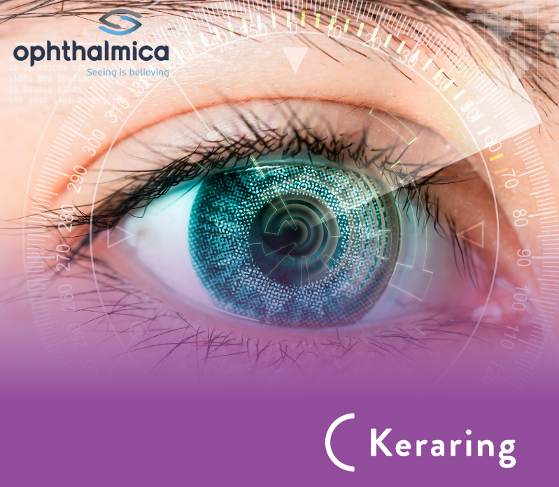
Ενδοστρωματικοί Κερατοειδικοί Δακτύλιοι Kerarings | Διόρθωση των διαθλαστικών ανωμαλιών που σχετίζονται με τον Κερατόκωνο
Οι Ενδοστρωματικοί Κερατοειδικοί Δακτύλιοι Kerarings είναι οφθαλμολογικά εμφυτεύματα που χρησιμοποιούνται για τη διόρθωση των διαθλαστικών ανωμαλιών που σχετίζονται με τον κερατόκωνο και άλλες εκτατικές διαταραχές του κερατοειδούς. Πρόκειται στην ουσία για δακτυλίους, οι οποίοι εμφυτεύονται στο εσωτερικό του οφθαλμού, και πιο συγκεκριμένα στο στρώμα του κερατοειδή, με σκοπό την αναδιαμόρφωση και ομαλοποίησή του σχήματός του. […]